ELBOW STIFFNESS/LIMITATION OF ELBOW MOVEMENTS
GENERAL INFORMATION
The function of the elbow joint is to provide the required position of the hand. Thus, the limitation of the elbow joint movements minimises the reaching area of the hand in space. The patient having the limitation of elbow movement may noteat, comh his hair or meet the toilet needs.
The hand should have and elbow joint with 30°-130° degrees of functional space to perform such daily activities. In other words, 100°’ elbow flexion/extansion movement is required. Besides, the pronation/supination move should also be approximately 100° (figure 1).
[auto_thumb width=”300″ height=”170″ link=”https://www.hakangundes.com.tr/wp-content/uploads/1n.png” lightbox=”true” align=”center” title=”” alt=”” iframe=”false” frame=”true” crop=”true”]https://www.hakangundes.com.tr/wp-content/uploads/1n.png[/auto_thumb]
WHAT IS THE CAUSE?
The movement limit in elbow joint is only one directional. This movement axis is called flexion/extension (opening/closing the elbow) (figure 1). In accordance with the features of each joint, either bone structure or soft tissues have more effect on the stability. The more the limit of movement a joint has, the more soft tissue support it needs for stability. For example, providing the stability of the shoulder joint is basically the mission of the adjacent soft tissues (joint capsule, ligaments, adjacent muscles-tendons). On the contrary, the limitation of movement (joint stiffness) is more commonly seen on such joints (figure 2). If a joint movement is one directional (i.e. only opening and closing of the elbow joint) and does not have many movement limits, less instability problems are observed on that joint. In summary, the elbow joint has more tendency to the limitation of movement, in other words the joint stiffness, more than the other joints.
[auto_thumb width=”300″ height=”170″ link=”https://www.hakangundes.com.tr/wp-content/uploads/2n.png” lightbox=”true” align=”center” title=”” alt=”” iframe=”false” frame=”true” crop=”true”]https://www.hakangundes.com.tr/wp-content/uploads/2n.png[/auto_thumb]
Figure 2. The elbow joint has more tendency to the limitation of movement, in other words the joint stiffness, more than the other joints
As the basic principle, the fractures around the elbow joint shoukd be duly fixed by surgical methods, the ligament and soft tissue injuries should be repaired and THE JOINT MOVEMENTS AND FUNCTIONS SHOULD BE RECOVERED IMMEDIATELY.
Before perfoming these, the union can be provided by keeping the elbow joint immobile (plaster etc.). ın this case, the elbow joint is also unioned together with the fractures and LIMITATION OF ELBOW MOVEMENTS occur. In other words, the union of the fractures does not mean anything since the joint will lose its functions. Before the surgical treatment, applying early movements passing the plaster treatment will cause some other problems. Since the fracture will not be unioned and the liagements will not be healed, INSTABILITY occurs. As the patient will have pain and pathological movements with each movement of the joint, the joint will lose its functions anyway.
In summary, the two main problems of the elbow joint, INSTABILITY AND LIMITATION OF MOVEMENT are observed on the patients who are not duly treated or never apply for the treatment.
The elbow stiffness is the most commonyl seen reason after the trauma. The studies have shwon that more than half of the patients have developed elbow joint stiffness after the elbow dislocations (or fractured dislocations).
CLASSIFICATION
The elbow joint stiffness is divided into three in the planning of the treatment:
1) Extrinsic: The thickhening and adhesion of the soft tissues (capsule, ligament, muscle) surrounding the elbow joint and prevention of the movement. There is no intraarticular problem and direct x-ray is normal (figure 3).
[auto_thumb width=”300″ height=”170″ link=”https://www.hakangundes.com.tr/wp-content/uploads/3n.png” lightbox=”true” align=”center” title=”” alt=”” iframe=”false” frame=”true” crop=”true”]https://www.hakangundes.com.tr/wp-content/uploads/3n.png[/auto_thumb]
Figure 3. Extrinsic: The thickhening and adhesion of the soft tissues (capsule, ligament, muscle) surrounding the elbow joint and prevention of the movement. Generally no bone pathology is detected in the direct x-ray.
2) Intrinsic: There is intraarticular problem. In other words, there is misunioned fractures, intraarticular adhesions, malalignment in the joint or joint cartilage loss. Direct x-ray is not normal (figure 4, 5, 6).
[auto_thumb width=”260″ height=”170″ link=”https://www.hakangundes.com.tr/wp-content/uploads/4n.png” lightbox=”true” align=”center” title=”” alt=”” iframe=”false” frame=”true” crop=”true”]https://www.hakangundes.com.tr/wp-content/uploads/4n.png[/auto_thumb]
[auto_thumb width=”300″ height=”170″ link=”https://www.hakangundes.com.tr/wp-content/uploads/5n.png” lightbox=”true” align=”center” title=”” alt=”” iframe=”false” frame=”true” crop=”true”]https://www.hakangundes.com.tr/wp-content/uploads/5n.png[/auto_thumb]
[auto_thumb width=”300″ height=”170″ link=”https://www.hakangundes.com.tr/wp-content/uploads/6n.png” lightbox=”true” align=”center” title=”” alt=”” iframe=”false” frame=”true” crop=”true”]https://www.hakangundes.com.tr/wp-content/uploads/6n.png[/auto_thumb]
Figure 4. Intrinsic: The fractures misunioned in wrong positions, joint cartilage loss etc. Direct x-ray is not normal.
Figure 5. Intrinsic: There is intraarticular problem. Intraarticular adhesion (osteophyte) is observed.
The Archive of Hakan Gündeş
Figure 6. Intrinsic: There is intraarticular problem. Joint cartilage loss is observed.
The Archive of Hakan Gündeş
Figure 7. Complex: In addition to the intraarticular problems, there is adhesion on the soft tissues around the joint, too. In other words, both intrinsic and extrinsic factors are combined.
The Archive of Hakan Gündeş
[auto_thumb width=”300″ height=”170″ link=”https://www.hakangundes.com.tr/wp-content/uploads/7n.png” lightbox=”true” align=”center” title=”” alt=”” iframe=”false” frame=”true” crop=”true”]https://www.hakangundes.com.tr/wp-content/uploads/7n.png[/auto_thumb]
3)Complex: The limitation starting with intrinsic factors at first is added the extrinsic factors as secondary dominant (secondary extrinsic). In other words, the pain occuring due to the intraarticular problems is tried to be treated by keeping the joint stabile and adhesion develops pn the soft tissues around the joint accordingly. In summary, both intrinsic and extrinsic factors are combined (figure 7).
DIAGNOSIS
In the diagnosing process, direct x-rays are required in addition to the medical history and physical examination. Under some conditions, CT is also required. In some stages of the disease, the body tries to cover the joint with a shield. This is called Heterotopic Ossification (figure 8). CT is required fort his case whose surgical plan and treatment is much harder. MRI examination is generally not necessary.
[auto_thumb width=”300″ height=”170″ link=”https://www.hakangundes.com.tr/wp-content/uploads/8n.png” lightbox=”true” align=”center” title=”” alt=”” iframe=”false” frame=”true” crop=”true”]https://www.hakangundes.com.tr/wp-content/uploads/8n.png[/auto_thumb]
Generally there is problem on the ulnar nerve which is passing through the elbow joint either due to the first trauma causing the joint stiffness or the joint stiffness itself. Thus, EMG called nerve conduction test should be made before the treatment. Likewise, the examination findings of this nerve should be recorded in detail (figure 9). Together with (preferrably) or after the surgical relaxation relaxation or mobilization surgery for the ulnar nerve should be performed (cubital tuınnel syndrome).
[auto_thumb width=”300″ height=”170″ link=”https://www.hakangundes.com.tr/wp-content/uploads/9n.png” lightbox=”true” align=”center” title=”” alt=”” iframe=”false” frame=”true” crop=”true”]https://www.hakangundes.com.tr/wp-content/uploads/9n.png[/auto_thumb]
DIFFERENTIAL DIAGNOSIS
If there is pain when the joint is stabile and not used, bone and bone infection should be eliminated as the reasons of limitation of movement.
In the pediatric patients having limitation of elbow movements, congenital reasons should be eliminated. The surgical treatment of such problems does generally not respond positively (figure 10).
[auto_thumb width=”300″ height=”170″ link=”https://www.hakangundes.com.tr/wp-content/uploads/10n.png” lightbox=”true” align=”center” title=”” alt=”” iframe=”false” frame=”true” crop=”true”]https://www.hakangundes.com.tr/wp-content/uploads/10n.png[/auto_thumb]
TREATMENT
Early movement is the most important part of the preventive treatment after the elbow trauma.
If there is fracture, this condition depends on the stability of the repair surgery performed. In the first group patients (extrinsic), rehabilitation can be performed until 6 months after the injury. Rehabilitation does not have any help in the second and third group (intrinsic) patients.
In the planning of the surgical treatment, the most important issue is the prevention of the limitation on the elbow on the daily life, in other words the patient should search for a treatment. Besides, the age and the functional needs of the patient should also be taken into consideration. The closing of the elbow is more important than the opening in the administration of daily life functions.
There is common opinion about the non-performance of surgery under the following conditions.
1-Surgery should not be performed for cosmetic reasons.
2-Surgery should not be performed if there is infection.
3-Surgery should not be performed if the circulation on the arm is ominous due to the perviously held trauma or surgical intervention.
4-The results are worse in pediatric patients.
5-The patient should be self-prepared for a long rehabilitation process and probable unsuccessful results.
The limitation of elbow joint movements is a persistent disease and requires a long term of struggle.
6-The surgery can be delayed if the loss on the ulnar nerve is severe.
SURGICAL TREATMENT
In the first group patient (extrinsic), the structures causing the adhesion and limitation of movement around the joint should be surgically removed. This surgery can be performed as open or arthroscopic surgery; or in combination (watch the videos in the operating theater).
Basically, even if the arthroscopic method is the preference of both the patient and the surgeon, open surgery results may be better in some cases. Your surgeon will explain you the reasons.
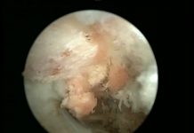
The movement limit acquired by artroscopic surgical relaxation is approximately 40 degrees. Since lens and optic camera is used in this method, intraarticular side willbe observed better, the pain will be less as the joint capsule is not cut and rehabilitation period will be easier (figure 11). But the disadvantages are that it cannot be applied for every patient since the acquired movement limit is less and there is potential nerve injury risk (radial and median nerve). In the second and third group patients (intrinsic), since there are anatomic disorders in the joint, the application chance is highly limited (figure 12). If the ulnar nerve is transferred to the fore side in the previous fracture repair surgery, arthroscopic methods should not be used.
[auto_thumb width=”350″ height=”199″ link=”https://www.hakangundes.com.tr/wp-content/uploads/11n.png” lightbox=”true” align=”center” title=”” alt=”” iframe=”false” frame=”true” crop=”true”]https://www.hakangundes.com.tr/wp-content/uploads/11n.png[/auto_thumb]
[auto_thumb width=”350″ height=”199″ link=”https://www.hakangundes.com.tr/wp-content/uploads/12n.png” lightbox=”true” align=”center” title=”” alt=”” iframe=”false” frame=”true” crop=”true”]https://www.hakangundes.com.tr/wp-content/uploads/12n.png[/auto_thumb]
In the open surgery method, all the structures causing adhesion and limitation of movement are removed by turning 360 degress around the joint. While performing this, the ligaments holding the joint should be kept stabile. If the ligaments should be cut to provide the movement, after the intervention is completed, the ligaments should be reconstructed (figure 13). Otherwise, an unstabile joint is acquired.
[auto_thumb width=”300″ height=”170″ link=”https://www.hakangundes.com.tr/wp-content/uploads/13n.png” lightbox=”true” align=”center” title=”” alt=”” iframe=”false” frame=”true” crop=”true”]https://www.hakangundes.com.tr/wp-content/uploads/13n.png[/auto_thumb]
In the open surgery method, the joint should be reached from exterior side, interior side or from both sides of the elbow joint (figure 14, 15, 16). The best approach for the patient is the approach that the surgeon feels more comfortable and easier to intervene the pathology (figure 17, figure 18 and figure 19). In the literature, the avarage movement gain is between 40-70 degrees (figure 20). There may be some loss in the acquired movement limit during the surgery in time. The movement acquired in the surgery cannot be increased with physical therapy and rehabilitation. Loss of movement acquired in the surgery is inevitable. Rehabilitation can help to minimize this loss.
[auto_thumb width=”260″ height=”170″ link=”https://www.hakangundes.com.tr/wp-content/uploads/14n.png” lightbox=”true” align=”center” title=”” alt=”” iframe=”false” frame=”true” crop=”true”]https://www.hakangundes.com.tr/wp-content/uploads/14n.png[/auto_thumb]
[auto_thumb width=”300″ height=”170″ link=”https://www.hakangundes.com.tr/wp-content/uploads/15n.png” lightbox=”true” align=”center” title=”” alt=”” iframe=”false” frame=”true” crop=”true”]https://www.hakangundes.com.tr/wp-content/uploads/15n.png[/auto_thumb]
[auto_thumb width=”300″ height=”170″ link=”https://www.hakangundes.com.tr/wp-content/uploads/16n.png” lightbox=”true” align=”center” title=”” alt=”” iframe=”false” frame=”true” crop=”true”]https://www.hakangundes.com.tr/wp-content/uploads/16n.png[/auto_thumb]
[auto_thumb width=”260″ height=”170″ link=”https://www.hakangundes.com.tr/wp-content/uploads/17n.png” lightbox=”true” align=”center” title=”” alt=”” iframe=”false” frame=”true” crop=”true”]https://www.hakangundes.com.tr/wp-content/uploads/17n.png[/auto_thumb]
[auto_thumb width=”300″ height=”170″ link=”https://www.hakangundes.com.tr/wp-content/uploads/18n.png” lightbox=”true” align=”center” title=”” alt=”” iframe=”false” frame=”true” crop=”true”]https://www.hakangundes.com.tr/wp-content/uploads/18n.png[/auto_thumb]
[auto_thumb width=”300″ height=”170″ link=”https://www.hakangundes.com.tr/wp-content/uploads/19n.png” lightbox=”true” align=”center” title=”” alt=”” iframe=”false” frame=”true” crop=”true”]https://www.hakangundes.com.tr/wp-content/uploads/19n.png[/auto_thumb]
[auto_thumb width=”300″ height=”170″ link=”https://www.hakangundes.com.tr/wp-content/uploads/20n.png” lightbox=”true” align=”center” title=”” alt=”” iframe=”false” frame=”true” crop=”true”]https://www.hakangundes.com.tr/wp-content/uploads/20n.png[/auto_thumb]
PROBABLE COMPLICATIONS
The surgical treatment of the elbow stiffness is difficult and inclined to have complications. The porbable complications in summary are:
•Nerve-vein injuries.
•Fracture formations.
•Infection.
•Hematoma.
•Compartment syndrome.
•Instability: Worse than elbow stiffness kötüdür.
•Recurrence of the stiffness and permanent pain.
•Progressive worsening of the ulnar nerve function.


