DESCRIPTION
The performance and feeling of our hands are provided by three different nerves. Respectively, these are radial, unlar and median nerves. There are some areas sensitive to pressure (compression) on the lines through which the nerves extends. Median nevre is generally subject to the increasing pressure (compression) on the palmar side of the wrist. The findings pccuring due to this pressure is called carpal tunnel syndrome.
There is a narrow tunnel through wihch the median nevre passes in the wrist. This tunnel is called carpal tunnel. The base of this tunnel is composed of the wrist bones while the roof is composed of a ligament called transverse carpalligament (figure 1). While 9 tendons are tghe median nevre proceeds from the forearm to the handi they pass through this tunnel in the wrist. The increase of the pressure or the constraction of the tunnel causes the compression of the median nevre.
[auto_thumb width=”320″ height=”160″ link=”https://www.hakangundes.com.tr/wp-content/uploads/1j1.jpg” lightbox=”true” align=”center” title=”” alt=”” iframe=”false” frame=”true” crop=”true”]https://www.hakangundes.com.tr/wp-content/uploads/1j1.jpg[/auto_thumb]
(Figure 1)
WHAT IS THE CAUSE?
No specific reason is found in most of the patients. The main pathology of these patients is the extreme thicknening of the ligament that the nevre passes under and the pressure it makes to the nevre. This case is called idiopathic carpal tunnel syndrome.
In some cases, even the ligament does not thicken too much, the undergoing median nevre shows extreme sensitivity due to some substructure problems of the body. Diabetics or some tyrodid gland diseases can be given as examples. Thus, carpal tunnel syndrome is more commonly seen in endocrine diseases like diabetics.
In some cases, it is seen that the thickness of the ligament passing over the nevre is normal but the median nevre is comopressed due to the extreme crowd in the tunnel (volume decrease). Osteoarthritis and rheumatoid arthritis are some examples. Likewise, in the rheumatoid arthritis disease, abnormal tenosynovitis is seen on the tissues surrounding the tendons passing through the tunnel, this causes to the crowd inside the tunnel and compression of the median nevre.
The hormonal changes seen during the pregnancy and brest-feeding period may result in the carpal tunnel disease by causing edema (water retention on the tissues) and volume decrease
HOW TO DIAGNOSE?
There are three main symptoms of the carpal tunnel sydrome. Pain, tingling and numbness. The tingling and numbness is felt on the thumb, forefinger and middle finger. It is not felt on the little (fifth) finger. The symptoms are generally seen during the nights as the wrist is bended in the state of sleep. The bended wrist decreases the volume in the carpal tunnel and increases the findings. The numbness and tingling generally wakes the patient up and he usually feels the need tos hake his hand. In the next steps, incompetence (i.e. while buttoning) dropping the thins from the hand, decrease in the strenght to grasp the things and myolysis on the atrophy around the thuBms can be observed due to the weakening of the effected muscles.
During the diagnosing period, the detailed medical history of the patient is studied. Even rarely, carpal tunnel syndrome might be the first findings of some endocrine and rheumatismal diseases. In this case, the required laboratory tests are to be made. In some arthritis-like diseases regarding the wrist, film mişght be required. To verify the diagnosis, generally a test called electromyography (EMG) is requested. In this test, the speed and health of the nevre conduction and other probable nevre compressions are researched. The compression may be observed on more than one area on the same nevre line (doublecrush). For example, median nevre can be compressed on both the wrist and the neck (cervical discal hernia). The test is performed by a neurologist or a physical therapist.
WHAT IS THE TREATMENT?
Upon establishing the diagnosis of the disease, the measures preventing the increase of pressure on the carpcal tunnel are taken. The wrist splint prescribed to use during the night will prevent the bending of the wrist and thus the pressure increase. If the patient has diabetics or rheumatism, first he should be examined by a specialist. These measures are generally sufficient at the early stage. In case of rheumatism etc, duly performed corticosteroid (cortisone) injection (needle) can regress the synovitis and edema. This injection can safely be made in the pregnancy and breast-feeding period.
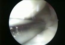
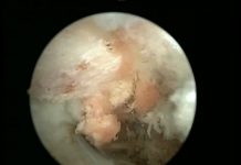
For the patients with unsuccesful results, surgical treatment is the only option. In the surgery, the transversecarpalligament passing over the nevre tissue is cut and carefully cleared of the adhesions. This surgery is generally performed with mini incision (figure 2). For the patients having rheumatism etc, the surgery should be performed with a standart big incision (figure 3). This incision is necessary to see all the pathologies and for the treatment (figure 4).
CERRAHİ TEDAVİ NASIL BİR SÜREÇ İZLER, BENİ NELER BEKLİYOR?
x-Ray graphy may be required after the consultation with the Orthopedician or Hand Surgeon. In most of the cases, daycase (short stay at the hospital) hospitalization is preferred. There is no need to apply general anesthesia. Generally, it is sufficient to make local (axiallry block or RIVA) anesthesia. During your consultation with your doctor, it is very important to mention about your special conditions (chronic diseases, regularly taken medication etc.). ın the early period after the surgery (postop 3 days), cold application and keeping your hand above the heart level will reileve the pain and throbbing. In the post op early period, your wrist will be covered by a bandage or a splint. The wound is controlled by opening the bandage between 5-7 days. If there is no complication, you can take shower. Even rarely, you might need physical therapy and rehabilitation after this period. Even though the general progress canges in accordance with the surgery performed and the condition of your wrist, you are generally expected to go bak your normal life within 3 weeks.
PROBABLE COMPLICATIONS
The most important complication is the injury of a branch (motor branch) located around the surgically operated area and originating from median nevre (figure 5). The operation should be performed under microscope. In case the nevre is injured, a long and troubled period starts.
Another probable complication is the insufficient opening of the ligament (sheath). In this case, the surgical process should be repeated. The secondary complications are limitation of finger movements due to tissue adhesion on the surgical wound area, chronic pain (RSD), gettin late or never gettin the expected results.
Carpal tunnel syndrome


